
There are two ways to define the time period that marks various milestones in fetal development: gestational and fertilization age.
These terms can be somewhat confusing since they are based upon different starting points. Fertilization age is a framework of time based upon the point of view of the unborn child. The gestational (or menstrual) age timeline begins two weeks earlier at the mother’s last menstrual period and is figured from her point of view. The framework most often used in discussions about the development of the unborn child is gestational age.
Thus, fertilization takes place at two weeks of gestational age and implantation about one week later. At fertilization, genetic instructions from the mother and the father combine to form a zygote, barely visible to the human eye.1 This single cell contains more information than fifty sets of the physical 33-volume set of the Encyclopedia Britannica.
On the very first day, the first four cell divisions take place as the zygote travels down the mother’s Fallopian tubes towards the uterus, all the while being nourished and protected by the mother’s body.
After 5 to 9 days, it implants in the uterus and from this point onward to about eight weeks is known as an embryo. Pro-abortionists often make the unscientific claim that pregnancy begins at implantation instead of fertilization. The change of terminology regarding the beginning of pregnancy was made in the 1960s based not on medical evidence, but simply to clear the way for abortifacient means of birth control that were under development at the time.2
Around 2 weeks after fertilization (four weeks gestational age), the first missed menstrual period happens. The mother’s menstruation is suppressed by chemical signals emitted by the unborn child. The child’s first completed brain cells appear.
At 3 weeks, the pre-born child’s heart is in an advanced stage of formation. His eyes begin to form, and his brain, spinal column, and nervous system are virtually complete.
At just over 3 weeks (24 days), his heart begins to beat [1].

At 4 weeks, his muscles are developing. His arm and leg buds are visible, and his first neocortical cells appear. The neocortex is the seat of complex thinking and reasoning, a feature present in no other mammal. He has grown in size by a factor of 10,000 since fertilization and is now 6 to 7 millimeters long (about ¼ inch). Blood flows in the baby’s veins, separate from his mother’s blood.
At 5 weeks, the baby’s pituitary gland is forming. The pituitary is often called the “master gland” since it controls the functions of most of the other endocrine glands. Additionally, the baby’s mouth, ears, and nose are taking shape.
At 6 weeks, the baby’s heart energy output is an incredible 20% that of an adult’s. Her cartilage skeleton is completely formed and ossification (bone formation) begins. The umbilical cord has developed. Her brain coordinates voluntary movement of muscles and the involuntary movement of organs. Reflex responses are present.
At around 43 days, the baby’s brain waves can be recorded.
At 45 days, he begins spontaneous and voluntary body movements, and his milk teeth buds are present.
At 7 weeks, his lips are sensitive to touch, and his ears resemble his family’s pattern. The first fully developed neurons (nerve cells) appear on the top of his spinal cord, beginning the construction of the brain stem, which regulates vital functions such as breathing, the heartbeat, and blood pressure.
At 8 weeks, the pre-born baby is well-proportioned, about 1½ inches long and 1/30 of an ounce in weight. All organs are present, complete, and functioning (except the lungs). Her heart beats sturdily. Her stomach produces digestive juices, her liver makes blood cells, and her kidneys are functioning. Her taste buds are forming and her unique fingerprints are being engraved. Her eyelids and the palms of her hands are sensitive to touch. Of the 45 total generations of cell replication that will take place by mature adulthood, fully two-thirds (30) have already taken place. She now consists of about one billion cells, containing more genetic information than every word communicated by every human being who has ever lived since the beginning of the human race.3
At 9 weeks, the pre-born child will bend his fingers around an object placed in his palm. His fingernails are forming and he sucks his thumb.
At 10 weeks, all sections of her body are sensitive to touch. She swallows, squints, frowns, and puckers up her brow.
By 11 weeks, he makes all facial expressions, including smiles. He is now breathing amniotic fluid steadily and will continue to do so until birth. His fingernails and toenails are now present. His taste buds are working; he will drink more amniotic fluid if it is artificially sweetened, and less if it is given a bitter taste.
By the end of the first trimester (12 weeks, the point at which most surgical abortions are done), vigorous activity shows the baby’s distinct personality. Sleep patterns differ; some babies hiccup constantly, others may cry. The baby can kick, turn over, curl and fan her toes, make a fist, open her mouth and press her lips tightly together, and practice breathing.
At 13 weeks, the preborn child’s facial expressions resemble those of her parents. Her movements are vigorous and graceful. Her vocal chords and external sex organs are present, and the sex of the baby can be determined. The baby can now hear very clearly.
At 4 months (16 weeks), the preborn baby can grasp with her hands, swim, and turn somersaults. Her mother may first feel her movements at this time. Her eyelashes are now present and rapid eye movements (REM), indicating dreaming, can be recorded. A very bright light shined on the mother’s abdomen will cause the baby to slowly move her arms to cover her eyes. Very loud music will cause her to cover her ears.
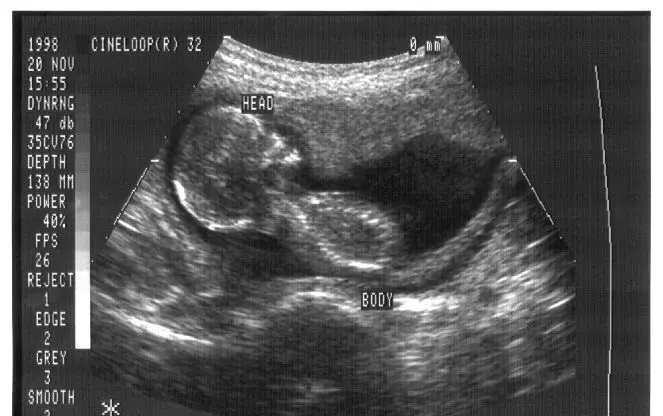
Ultrasound at four months.
By 5 months (20 weeks), the preborn baby has formed his own unique sleeping habits, and a loud sound such as a slammed door may startle him. His hearing has a wider range of frequency than an adult’s in both the higher and lower ranges. He may be soothed to sleep by gentle music.
At 6 months (24 weeks), most babies can live outside the womb with proper care. Fine hair grows on his head and eyebrows and he now has eyelashes. He weighs about 22 ounces and is about nine inches tall.
At 7 months (28 weeks), his weight increases to over one kilogram (2.2 pounds). His lungs are capable of breathing air. His eyeteeth are present. His hands can support his entire weight at this time, and he recognizes his mother’s voice. Of the 45 total generations of cell replication that will take place by mature adulthood, 38 have already taken place. He now has about 300 billion cells.
At 8 months (32 weeks), with only one month to go, his weight is about two kilograms (4.4 pounds). If born now, he has more than a 90% chance of surviving and being entirely healthy.
At 9 months (36 weeks), the baby is gaining about an ounce of weight per day. All of his senses are fully functioning, and his toenails and fingernails are complete.
On her birthday, the baby releases hormones that trigger labor. The lightest baby ever born to survive in good health weighed in at a mere 9.2 ounces, less than a can of soft drink. Of the 45 total generations of cell replication that will take place by mature adulthood, 41 have taken place by birth. The baby now has about two trillion (2,000,000,000,000) cells. The remaining four generations of cell replication will occupy all of the person’s childhood and young adulthood. This means, that, in developmental terms, we spend more than 90% of our lives in utero.
Fetal Development and the Pro-Life Cause
We should all be familiar with our own prenatal histories, not just because it is so fascinating, but because we might save an unborn child ourselves one day with this timeline of fetal development.
The greatest pro-life visual tools are not photos of aborted late-term pre-born babies, although these are indeed very powerful images. The most powerful persuaders are the full-color, beautiful, clear ultrasounds of pre-born babies peacefully floating in their mother’s wombs.

3D ultrasound at about 21 weeks.
The pro-abortionists clearly know this. When pro-lifers put up posters or billboards depicting living unborn children in a public place, abortion advocates often loudly object and even vandalize the displays. Also, in those rare instances when abortion advocates debate pro-lifers, they try their best to censor such images of ultrasounds. For example, third-trimester abortionist Warren Hern has written, “We respond to all requests from schools for educational presentations concerning abortion. If the sponsors want both sides presented, however, the presentations must be made on different occasions. We insist that visual aid materials not be presented by either side.”4
Regarding ultrasonography, pro-abortionist Sarah Ackley said [2], “The anti-abortion movement can, with little effort, marshal such images as evidence of fetal personhood. With no comparably powerful set of imagery at its disposal thus far, the abortion-rights movement will likely remain on the defensive with respect to questions of fetal personhood.”
Preborn children can be the pro-life movement’s most eloquent messengers ― but only if we are their voice.
+ Endnotes
[1] All information on fetal development is from the most-used medical dictionary in the world: Donald Venes, M.D. (Editor). Taber’s Cyclopedic Medical Dictionary. 2021: 24th Edition. F.A. Davis Company.
[2] In 1963, the generally accepted definition of “abortion” was “all the measures which impair the viability of the zygote at any time between the instant of fertilization and the completion of labor” (Public Health Service leaflet No. 1066, United States Department of Health, Education and Welfare [HEW], 1963, page 27). Two years later, the definition of the beginning of pregnancy had magically changed to “the implantation of a fertilized ovum” (American College of Obstetrics and Gynecology [ACOG]. Terminology Bulletin, “Terms Used in Reference to the Fetus” [Chicago: ACOG], September 1965).
[3] The total amount of information communicated since Adam and Eve were created has been contained in about 12 billion hardback and softback volumes averaging 180 pages and 55,000 words apiece; about 500 billion separate copies of newspapers, magazines, periodicals and newsletters of every description, each averaging 45 pages and 25,000 words; and about 100 billion people having spoken an average of 7,500 words per day for an average 45‑year lifespan. This is a total of 12,331,910,000,000,000,000 words of communication transmitted in every form since human life began on Earth (give or take a few quadrillion).
[4] Warren Hern, M.D. Abortion Practice (New York City: B. Lippincott Company), 1984, page 323.